Last updated: January 9, 2026
Any links on this page that lead to products on Amazon are affiliate links and I earn a commission if you make a purchase. Thanks in advance – I really appreciate it!
CALL YOUR VETERINARIAN IMMEDIATELY IF:
- Bleeding soaks through a firmly-held towel within 5 minutes (indicates possible arterial involvement).
- You see exposed white/silver tissue (visible tendon, ligament, or bone).
- Puncture wound exceeds ½ inch deep or is located near a joint or tendon sheath.
- Joint involvement is suspected: Heat, massive swelling, or thick, clear “joint oil” leaking from a wound.
- Severe lameness (Grade 3+): Horse is “toe-touching” or refusing to bear weight.
- Wound is on the eyelid, chest, or over vital organs.
- Obvious contamination: Deeply embedded wood, metal, wire, or soil.
- Signs of shock: Pale gums, rapid breathing, weak pulse, or profuse sweating.
Proper horse wound care starts with split-second decisions that mean the difference between a barrel horse that competes again and one with permanent tendon damage. After 30+ years managing wounds on my horses—from pasture barbed wire cuts to racetrack kick wounds—I’ve learned that knowing when to call the vet matters as much as what you do while waiting.
This guide walks you through the exact emergency triage system I use: how to assess bleeding severity, identify deep structure involvement, and perform safe first-aid without compromising veterinary treatment. You’ll learn the 5-minute bleeding test, the “joint poke” rule, and why that innocent-looking puncture wound near the fetlock demands immediate attention.
About This Guide: Written by Miles Henry, Louisiana-licensed racehorse owner (License #67012) with 30+ years of hands-on experience managing training and racing injuries. This information supports—but does NOT replace—veterinary diagnosis and treatment. Always consult your equine veterinarian for wound assessment.

Table of Contents
Understanding Wound Severity: The Triage System
Not all wounds require emergency veterinary care, but distinguishing between “watch closely” and “call now” requires systematic assessment. Here’s the triage framework veterinarians use—and what I apply during that critical first evaluation.
The 4 Critical Assessment Factors
1. Bleeding Pattern & Volume: Arterial bleeding (bright red, pulsing spurts) versus venous bleeding (dark red, steady flow) versus capillary oozing. If direct pressure doesn’t slow bleeding within about 5 minutes, more serious vessel involvement is likely. Veterinary wound-care references emphasize controlling bleeding with direct pressure and assessing the pattern and volume of blood loss.
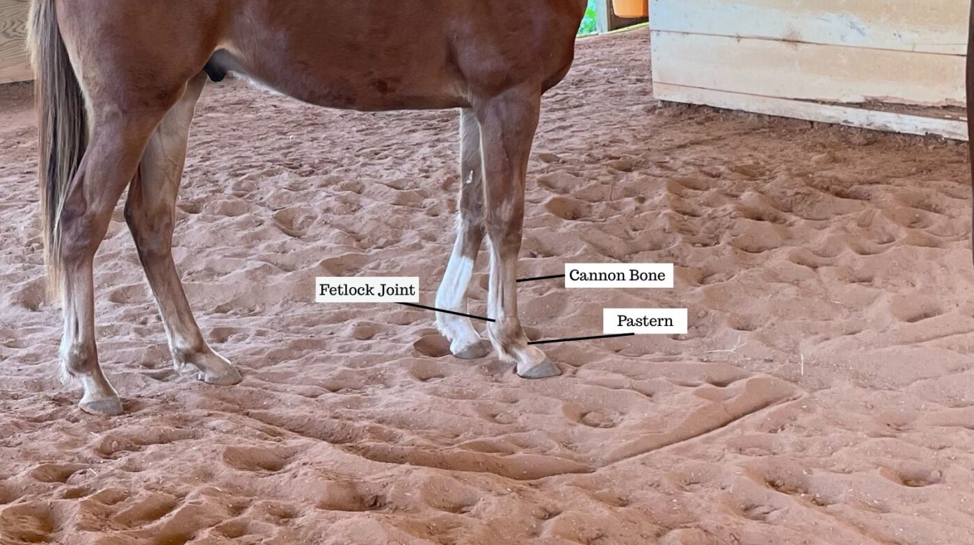
2. Wound Depth & Structure Involvement: Superficial wounds affect only the skin. Deep wounds expose or damage underlying tendons, ligaments, joint capsules, or bone. The “white tissue test”—if you see glistening white or silver strands, you may be looking at tendon or ligament.
3. Location Relative to Vital Structures: Wounds within 2 inches of joints, tendon sheaths, or major vessels carry a higher infection and complication risk. Lower limb wounds (below the knee/hock) heal more slowly due to reduced blood supply and constant motion.
4. Contamination Level: Clean wounds (smooth metal, glass) versus contaminated wounds (barbed wire, wood) versus grossly contaminated wounds (farm equipment, soil, manure). Research on contaminated wounds shows that bacterial load and type of contamination significantly affect infection risk and management strategies.
| Wound Type | Bleeding Pattern | Depth/Structures | Key Identifying Signs | Veterinary Urgency |
|---|---|---|---|---|
| Minor Laceration | Slow oozing; stops with direct pressure. | Skin only; | Clean edges, minimal gaping, horse is full weight-bearing. | Low: Within 24 hours (for cleaning/stitching). |
| Infected Puncture | Minimal; often appears dry or “crusted.” | Deep, narrow track; risk of bone/joint infection. | Sudden heat, swelling, pus, or “3-legged” lameness. | IMMEDIATE: Risk of sepsis or tetanus. |
| Tendon/Ligament | Moderate to heavy. | Exposed white “cord-like” tissue. | Abnormal limb angle or dropped fetlock; severe lameness. | EMERGENCY: Minimize movement, call vet now. |
| Joint Penetration | Variable; may be minimal. | Into joint capsule or tendon sheath. | Thick, clear, oily fluid (synovial fluid) leaking from hole. | CRITICAL: Must be flushed within 6–8 hours. |

Safe First-Aid Actions While Awaiting Veterinary Care
The 30–60 minutes before your veterinarian arrives are critical. Well-intentioned first-aid, like applying harsh chemicals or heavy ointments, can actually damage healthy tissue and interfere with suturing. Follow these vet-approved steps to stabilize your horse safely.
VET-APPROVED OWNER ACTIONS
- DIRECT PRESSURE: Use a clean towel. Hold for 5 minutes without “peeking.” If it soaks through, add another layer—never remove the bottom towel.
- SALINE FLUSH: Use 0.9% sterile saline only. (Mix 1 tsp non-iodized salt into 2 cups of boiled-then-cooled water if you are out of medical saline).
- STALL REST: Confine the horse immediately to a small, clean area to prevent movement from reopening vessels or driving dirt deeper.
- PHOTO TRIAGE: Take clear, well-lit photos of the wound *before* wrapping to send to your vet for assessment.
Once bleeding is controlled with pressure, you can further stabilize the injury by encouraging the horse to rest the limb slightly raised. Cover the area with a non-adherent pad (like Telfa) and a loose wrap to keep dirt out. Most importantly, check your records for the last Tetanus booster; AAEP guidelines suggest a booster for any contaminated wound if the last dose was more than 6 months ago.
❌ Common First-Aid Mistakes (Never Do These)
Using Hydrogen Peroxide or Betadine Scrub in the Wound: Both can damage healthy tissue needed for healing. Hydrogen peroxide foam may drive bacteria deeper. Diluted Betadine solution is sometimes used on intact skin around a wound, but the wound bed itself is usually better managed with saline unless your vet directs otherwise. University of Minnesota Extension
Applying Heavy Ointments Before Vet Evaluation: Thick antibiotic ointments can trap bacteria, make it difficult to assess the wound, and interfere with suturing. Wait until your veterinarian recommends a specific topical.
Attempting to “Stitch” or Close Wounds: Only veterinarians should close wounds. Some wounds should never be closed, and closure after too long can trap infection. Timing and wound type matter.
Administering Oral Antibiotics Without Vet Approval: Wrong drug, dose, or duration can worsen resistance and hide important signs. Your vet will select the right medication, route, and schedule.
Removing Embedded Foreign Objects: Deeply embedded posts, wire, or metal may be tamponading (plugging) major vessels. Removing them in the field can trigger massive bleeding. Stabilize the object if possible and let your veterinarian remove it under controlled conditions.
Using Tourniquets: Tourniquets are almost never appropriate for horse limb wounds outside a hospital setting. Direct pressure controls the vast majority of bleeding without risking tissue death from prolonged occlusion.
🔵 DAILY WOUND MONITORING (DURING HEALING)
THE 5-POINT MORNING CHECK
Monitoring these parameters during your daily grooming routine is the best way to catch complications early. While mild swelling peaks around day three, any progressive heat or foul-smelling, yellow discharge suggests an infection. Watch for the “white-pink” transition: healthy granulation tissue should look like moist, pink cobblestones. If it becomes dark, mushy, or raised (proud flesh), or if the horse develops a fever above 101.5°F, call your vet immediately.
RED FLAG: If any of these parameters worsen, or if your horse develops a fever (rectal temperature above about 101.5°F), contact your veterinarian the same day.
From the Track: Real Wound Cases & Lessons Learned
Theory meets reality when you’re standing in a stall at 5:30 AM with a bleeding horse. These cases from my racetrack experience taught me hard lessons about wound assessment, owner limits, and why “waiting to see” can backfire.
Case 1: The “Innocent” Puncture That Wasn’t
The Scenario: A three-year-old filly came in from turnout with a pencil-sized puncture wound on her right front fetlock. Minimal bleeding, barely limping. I cleaned it with saline, applied a light wrap, and planned to call the vet “if it got worse.”
What Happened: By evening feeding (about 6 hours later), the fetlock had swollen to twice its normal size, was hot, and the filly was Grade 4 lame (toe-touching only). The vet aspirated the joint, positive for synovial fluid. The puncture had penetrated the fetlock joint capsule.
The Lesson: Any puncture wound within about 2 inches of a joint gets immediate veterinary evaluation, no exceptions. Joint infections often need lavage and antibiotics within hours. We were on the late side but still in time to save the joint; she recovered after hospital care, but earlier evaluation would have been safer and less expensive.
Case 2: Wire Laceration—When Size Doesn’t Tell the Story
The Scenario: A gelding got tangled in old barbed wire during a storm. I noticed he had about a 6-inch laceration across the gaskin, with moderate bleeding controlled by pressure. The wound looked “clean” with smooth edges—classic wire cut.
The Decision: I called the vet immediately, despite it being late at night. The location (lower limb), length (likely needed sutures), and contamination (rusty wire and mud) were non-negotiable red flags.
The Outcome: After clipping and cleaning, the vet found a small wire barb embedded under the skin flap, invisible from the surface. It had tracked along the muscle sheath. Removing it under sedation and sterile conditions prevented a deep-tissue abscess.
The Lesson: Barbed wire injuries always get immediate veterinary care—no exceptions. Wire can create hidden pockets and embedded fragments that aren’t visible from the outside.
Case 3: The Proud Flesh Problem—Prevention vs. Treatment
The Scenario: A mare sustained a 3-inch laceration on her left hind cannon bone. The wound was sutured within 4 hours, and bandage care was excellent. No early infection. By week three, instead of closing, the wound developed exuberant granulation tissue (proud flesh) that grew above skin level and prevented closure.
The Treatment: She required weeks of repeated treatments to control the granulation tissue and allow skin to grow across it. Healing took much longer than originally expected.
The Lesson: Lower limb wounds, especially on the front of the cannon bone, are prone to proud flesh. Today, I discuss early options like skin grafts or advanced therapies with my vet if a lower-leg wound isn’t closing on schedule; prevention is easier than long-term management.
Case 4: When “Wait and See” Was Correct
The Scenario: I had a four-year-old colt suffer a superficial scrape across his shoulder about 4 inches long and 1 inch wide. The skin was irritated but intact in deeper layers. He was sound at trot, with a full range of motion.
The Decision: I cleaned it with saline, applied a non-stick dressing for a few days, and monitored twice daily. No vet call—just careful observation and documentation.
The Outcome: The wound formed a healthy scab by day three, began new skin growth by day seven, and fully healed by week five with no complications.
The Lesson: Not every wound requires immediate veterinary intervention. Location (upper body vs. lower limb), depth, cleanliness, and the horse’s comfort level all matter. If that same wound had been on his lower leg instead of his shoulder, I would have called the vet due to the higher risk of proud flesh and slower healing.
Wound Treatment Phases: What to Expect
Phase 1: Inflammatory Phase (0–5 Days)
What’s Happening: Blood clotting seals the wound, and white blood cells move in to fight bacteria. The wound often looks red, swollen, and warm—this can be normal early in healing.
Normal Signs: Moderate swelling that peaks around day 2–3 and then holds or begins to improve, clear to slightly pink drainage, firm clot formation, mild to moderate pain or lameness.
Concerning Signs: Swelling that continues to increase after day 3, yellow/green discharge, severe pain, fever above about 101.5°F, or red streaks tracking up the limb (lymphangitis).
Phase 2: Proliferative Phase (5 Days – 3 Weeks)
What’s Happening: Granulation tissue fills the wound, new blood vessels form, and the wound begins to contract as edges are pulled together.
Normal Signs: Pink to red granulation tissue with a moist, “cobblestone” appearance, decreasing drainage, wound edges gradually pulling together, improving lameness.
Concerning Signs: Granulation tissue growing above skin level (proud flesh), pale or dark granulation tissue, lack of contraction, persistent heavy drainage, or little change in lameness.
Phase 3: Remodeling Phase (3 Weeks – Many Months)
What’s Happening: New skin cells cover granulation tissue, collagen reorganizes to strengthen the wound, and the scar matures.
Normal Signs: Thin pink skin growing inward from wound edges, smoother surface over time, white hair growth in the scar, gradual return to soundness.
Concerning Signs: Wound reopening (dehiscence), excessive scar tissue that interferes with movement, chronic drainage, or lameness that persists or worsens. Chronic lameness after wound healing often requires farrier/vet re-evaluation.
Special Wound Scenarios: Joint, Tendon & Eye Injuries
Suspected Joint Penetration
Joint infections (septic arthritis) can be career-ending if not treated within hours. Early signs include:
- Wound located within roughly 2 inches of a joint (fetlock, knee, hock, stifle, shoulder).
- Clear, sticky “joint oil” fluid leaking from the wound.
- Rapid joint swelling over a few hours.
- Severe lameness that seems out of proportion to wound size.
- Joint feels hot compared to the opposite limb.
Real Case (Track Experience): I had a racehorse who hit her legs every race due to repeated overreach trauma. What looked like bumps soon affected her joints and at-speed mechanics, and ultimately, we had to take her off the track. That experience taught me the importance of early intervention and correct trimming, booting, and turnout strategies. For a full breakdown of prevention and treatment, see our overreach injuries guide.
Veterinary Assessment: Your veterinarian may perform a sterile fluid injection test into the joint and watch whether it exits through the wound, confirming communication. This is not something to attempt yourself.
Tendon & Ligament Lacerations
Exposed tendons or ligaments often appear as glistening white or silver bands with visible fiber pattern. These injuries are most common where tendons are closest to the skin, such as the back of the cannon bone.
Emergency Protocol: Cover the area promptly with saline-moistened sterile gauze and a protective wrap to prevent the tissue from drying out. Call your veterinarian immediately for evaluation and a surgical plan if needed.
Eyelid & Eye Area Wounds
Eye and eyelid injuries are always urgent. Eyelids are crucial for protecting the eye, and even small lid wounds need expert repair to preserve function and prevent corneal exposure.
Safe First Steps: Place the horse in a quiet, dim area if possible. Gently cover the eye with a clean, damp cloth and avoid applying any medications unless your veterinarian has instructed you to do so. Add eye checks to your daily grooming routine.
Preventing Common Wound Scenarios
Turnout & Fencing Safety
Many serious lacerations are fence- or gate-related. Barbed wire, loose woven wire, and broken boards are frequent culprits. Regular inspections and timely repairs dramatically reduce injury risk.
Walk your pastures and pens looking for:
- Barbed wire or old wire lying in the grass.
- Broken boards or splintered rails.
- Exposed nail or screw tips.
- Sharp gate hardware and protruding bolts.
Tetanus Protection & Vaccination
Tetanus is a nearly always fatal disease in horses, but it is highly preventable with proper vaccination. Because Clostridium tetani bacteria live in soil and manure, veterinarians consider tetanus protection essential for all horses.
Work with your veterinarian to ensure your vaccination program follows current recommendations and to decide when a booster is appropriate after a wound.
- Horses on a standard vaccination schedule often receive tetanus annually.
- If a wound occurs and it has been more than about 6 months since the last tetanus shot, your vet may recommend a booster within 24 hours.
- Unvaccinated horses or those with unknown status may need both tetanus toxoid and antitoxin.
Essential Wound Care Kit
Having the right supplies ready can save precious minutes when a wound happens. A basic barn kit might include:
- Sterile saline solution and large syringes for gentle flushing.
- Sterile gauze sponges and non-stick dressings.
- Padding (sheet cotton or quilted wraps) and cohesive bandages.
- Bandage scissors, disposable gloves, and a thermometer.
- Your veterinarian’s phone number clearly posted in the barn.
Important: This guide is designed to help you recognize emergencies quickly and provide safe first-aid until your veterinarian can take over. It is not a substitute for professional diagnosis or treatment. When in doubt—even if the wound looks “small”—contact your veterinarian early. In 30+ years, the only regret seen is waiting too long, not calling too soon.
Dr. Erica Lacher from Springhill Equine Veterinary Clinic demonstrates the exact first 10 minutes of wound care every horse owner should know before the vet arrives.

Frequently Asked Questions About Horse Wound Care.
Can you use hydrogen peroxide on horse wounds?
Hydrogen peroxide is not recommended for treating wounds on horses. It can damage healthy tissue, delay healing, and may even drive bacteria deeper into the wound. Most veterinarians prefer gentle flushing with sterile saline unless they advise otherwise.
Why do wounds on horses result in proud flesh?
Proud flesh results from excessive granulation tissue during healing, especially on lower limbs where there is less blood supply and more motion. Factors such as infection, constant movement, and inadequate bandaging increase the risk. Management usually involves careful bandaging and veterinary-guided treatments to control the excess tissue.
Not sure if a cut requires a vet? Check our Horse First Aid Emergency Protocol to see if your horse’s vitals are in the danger zone.
About Miles Henry
Racehorse Owner & Author | 30+ Years in Thoroughbred Racing
Miles Henry (legal name: William Bradley) is a professional horseman based in Folsom, Louisiana. He holds Louisiana Racing License #67012 and has spent over three decades managing Thoroughbreds at premier tracks including Fair Grounds, Delta Downs, and Evangeline Downs.
Expertise & Hands-On Experience: Beyond the track, Miles has decades of experience in specialized equine care, covering everything from hoof health and nutrition to training protocols for Quarter Horses, Friesians, and Paints. Every guide on Horse Racing Sense is rooted in this “boots-on-the-ground” perspective.
30 of their last 90 starts
Equibase Profile.
Connect with Miles: